Health Topics
Myocardial biopsy is the removal of a small piece of heart muscle for examination.
How the Test is Performed
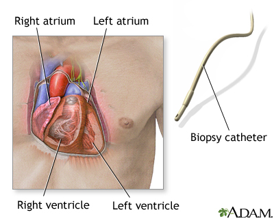
Myocardial biopsy is done through a catheter that is threaded into your heart (cardiac catheterization). The procedure will take place in a hospital radiology department, special procedures room, or cardiac diagnostics laboratory.
To have the procedure:
- You may be given medicine to help you relax (sedative) before the procedure. However, you will remain awake and able to follow instructions during the test.
- You will lie flat on a stretcher or table while the test is being done.
- The skin is scrubbed and a local numbing medicine (anesthetic) is given.
- The doctor inserts a thin tube (catheter) through a vein or artery, depending on whether tissue will be taken from the right or left side of the heart.
- If the biopsy is done without another procedure, the catheter is most often placed through a vein in the neck and then carefully threaded into the heart. The doctor will use moving x-ray images (fluoroscopy) or echocardiography (ultrasound) to guide the catheter to the correct area.
- Once the catheter is in position, a special device with small jaws on the tip is used to remove small pieces of tissue from the heart muscle.
- The procedure may last 1 or more hours.

How to Prepare for the Test
You will be told not to eat or drink anything for 6 to 8 hours before the test. The procedure takes place in the hospital. Most often, you will be admitted the morning of the procedure, but in some cases, you may need to be admitted the night before.
A health care provider will explain the procedure and its risks. You must sign a consent form.
How the Test will Feel
You may feel some pressure at the biopsy site. You may have some discomfort due to lying still for a long period of time.
Why the Test is Performed
This procedure is routinely done after heart transplantation to watch for signs of rejection.
Your provider may also order this procedure if you have signs of:
- Alcoholic cardiomyopathy
- Cardiac amyloidosis
- Cardiomyopathy
- Hypertrophic cardiomyopathy
- Idiopathic cardiomyopathy
- Ischemic cardiomyopathy
- Myocarditis
- Peripartum cardiomyopathy
- Restrictive cardiomyopathy
Normal Results
A normal result means no abnormal heart muscle tissue was discovered. However, it does not necessarily mean your heart is normal because sometimes the biopsy can miss abnormal tissue.
What Abnormal Results Mean
An abnormal result means abnormal tissue was found. This test may reveal the cause of cardiomyopathy. Abnormal tissue may be due to:
- Amyloidosis
- Myocarditis
- Sarcoidosis
- Transplant (heart) rejection
Risks
Risks are moderate and include:
- Blood clots
- Bleeding from the biopsy site
- Cardiac arrhythmias
- Infection
- Injury to the recurrent laryngeal nerve
- Injury to the vein or artery
- Pneumothorax
- Rupture of the heart (very rare)
- Tricuspid regurgitation
Alternative Names
Heart biopsy; Biopsy - heart
References
Kliger CA, Sorajja P. Special techniques. In: Sorajja P, Lim MJ, Kern MJ, eds. Kern's Cardiac Catheterization Handbook. 7th ed. Philadelphia, PA: Elsevier; 2020:chap 9.
Miller DV. Cardiovascular system. In: Goldblum JR, Lamps LW, McKenney JK, Myers JL, eds. Rosai and Ackerman's Surgical Pathology. 11th ed. Philadelphia, PA: Elsevier; 2018:chap 42.
Rogers JG, O'Connor CM. Heart failure: epidemiology, pathobiology, and diagnosis. In: Goldman L, Cooney KA, eds. Goldman-Cecil Medicine. 27th ed. Philadelphia, PA: Elsevier; 2024:chap 45.
Review Date 5/8/2024
Updated by: Thomas S. Metkus, MD, Assistant Professor of Medicine and Surgery, Johns Hopkins University School of Medicine, Baltimore, MD. Also reviewed by David C. Dugdale, MD, Medical Director, Brenda Conaway, Editorial Director, and the A.D.A.M. Editorial team.


